


Home / Training / Manuals / Atlas of breast cancer early detection / Cases
Atlas of breast cancer early detection
Go back to the list of case studies
.png) Click on the pictures to magnify and display the legends
Click on the pictures to magnify and display the legends
| Case number: | 041 |
| Age: | 64 |
| Clinical presentation: | Postmenopausal woman with increased risk of developing breast cancer because of breast carcinoma in her daughter presented for screening for breast cancer. Examination revealed a lump, 1.5 cm in diameter, in the upper outer quadrant of the left breast. |
Mammography:
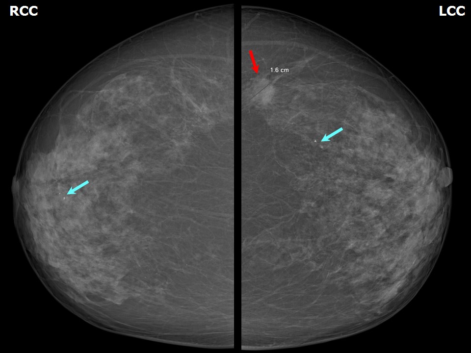 |  |
| Breast composition: | ACR category c (the breasts are heterogeneously dense, which may obscure small masses) | Mammography features: |
| ‣ Location of the lesion: | Left breast, upper outer quadrant at 23 oclock, posterior third |
| ‣ Mass: | |
| • Number: | 1 |
| • Size: | 1.8 × 1.6 × 1.4 cm |
| • Shape: | Irregular |
| • Margins: | Microlobulated |
| • Density: | Equal |
| ‣ Calcifications: | |
| • Typically benign: | None |
| • Suspicious: | None |
| • Distribution: | None |
| ‣ Architectural distortion: | None |
| ‣ Asymmetry: | None |
| ‣ Intramammary node: | None |
| ‣ Skin lesion: | None |
| ‣ Solitary dilated duct: | None |
| ‣ Associated features: | None |
| Breast composition: | ACR category c (the breasts are heterogeneously dense, which may obscure small masses) | Mammography features: |
| ‣ Location of the lesion: | Bilateral breasts: Right: upper inner quadrant at 1 o'clock, anterior third. Left: upper outer quadrant at 1 o'clock, middle third |
| ‣ Mass: | |
| • Number: | 0 |
| • Size: | None |
| • Shape: | None |
| • Margins: | None |
| • Density: | None |
| ‣ Calcifications: | |
| • Typically benign: | Round |
| • Suspicious: | None |
| • Distribution: | None |
| ‣ Architectural distortion: | None |
| ‣ Asymmetry: | None |
| ‣ Intramammary node: | None |
| ‣ Skin lesion: | None |
| ‣ Solitary dilated duct: | None |
| ‣ Associated features: | None |
Ultrasound:
 |  |
| Ultrasound features: Left breast, outer quadrants at 3 oclock position | |
| ‣ Mass | |
| • Location: | Left breast, outer quadrants at 3 oclock position |
| • Number: | 1 |
| • Size: | 1.1 × 1.0 cm |
| • Shape: | Irregular |
| • Orientation: | Not parallel |
| • Margins: | Spiculated |
| • Echo pattern: | Hypoechoic |
| • Posterior features: | Posterior shadowing |
| ‣ Calcifications: | None |
| ‣ Associated features: | None |
| ‣ Special cases: | None |
BI-RADS:
BI-RADS Category: 5 (highly suggestive of malignancy)Further assessment:
Further assessment advised: Referral for cytologyCytology:
 |
| Cytology features: | |
| ‣ Type of sample: | FNAC |
| ‣ Site of biopsy: | |
| • Laterality: | Left |
| • Quadrant: | Upper outer |
| • Localization technique: | Palpation |
| • Nature of aspirate: | whitish |
| ‣ Cytological description: | Smears are very cellular with many dyscohesive clusters of malignant ductal cells. Many single isolated malignant cells are also seen |
| ‣ Reporting category: | Malignant |
| ‣ Diagnosis: | Carcinoma low grade |
| ‣ Comments: | None |
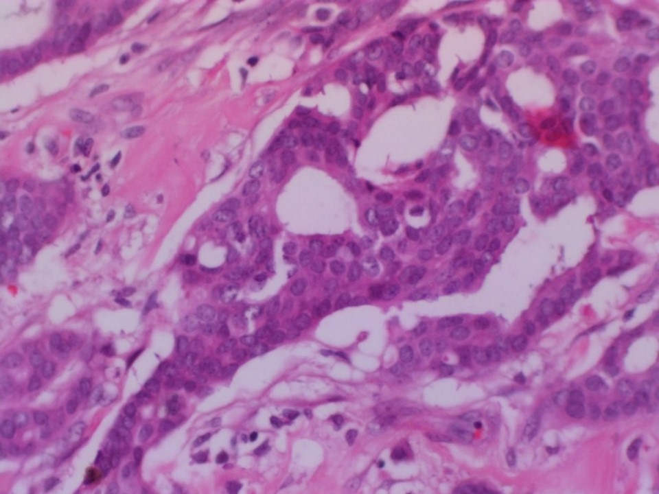
Histopathology:
MRM
 |
| Histopathology features: | |
| ‣ Specimen type: | MRM |
| ‣ Laterality: | Left |
| ‣ Macroscopy: | Cut surface shows a tumour (2.0 × 1.5 × 1.5 cm) in the lateral quadrant. Tumour is 1.5 cm from the base and 3.5 cm from the skin. Cut surface of tumour is firm to hard in consistency, greyish white with infiltrating margins. Areas of fibrosis are seen in the remaining breast |
| ‣ Histological type: | Invasive cribriform carcinoma |
| ‣ Histological grade: | Grade 1 (1 + 1 + 1 = 3) |
| ‣ Mitosis: | 2 |
| ‣ Maximum invasive tumour size: | 2.0 cm in greatest dimension |
| ‣ Lymph node status: | 0/14 |
| ‣ Peritumoural lymphovascular invasion: | Present |
| ‣ DCIS/EIC: | Cribriform DCIS of low grade |
| ‣ Margins: | Free of tumour, distance of tumour from the margin is 1.5 cm |
| ‣ Pathological stage: | pT1N0 |
| ‣ Biomarkers: | ER positive, PR positive, and HER2/neu negative |
| ‣ Comments: | Surrounding breast shows fibrosis and cyst formation |
Case summary:
| Postmenopausal woman presented with left breast lump. Diagnosed as left breast carcinoma, BI-RADS 5 on imaging, as breast carcinoma on cytology, and as invasive cribriform carcinoma, pT1N0 on histopathology. |
Learning points:
|